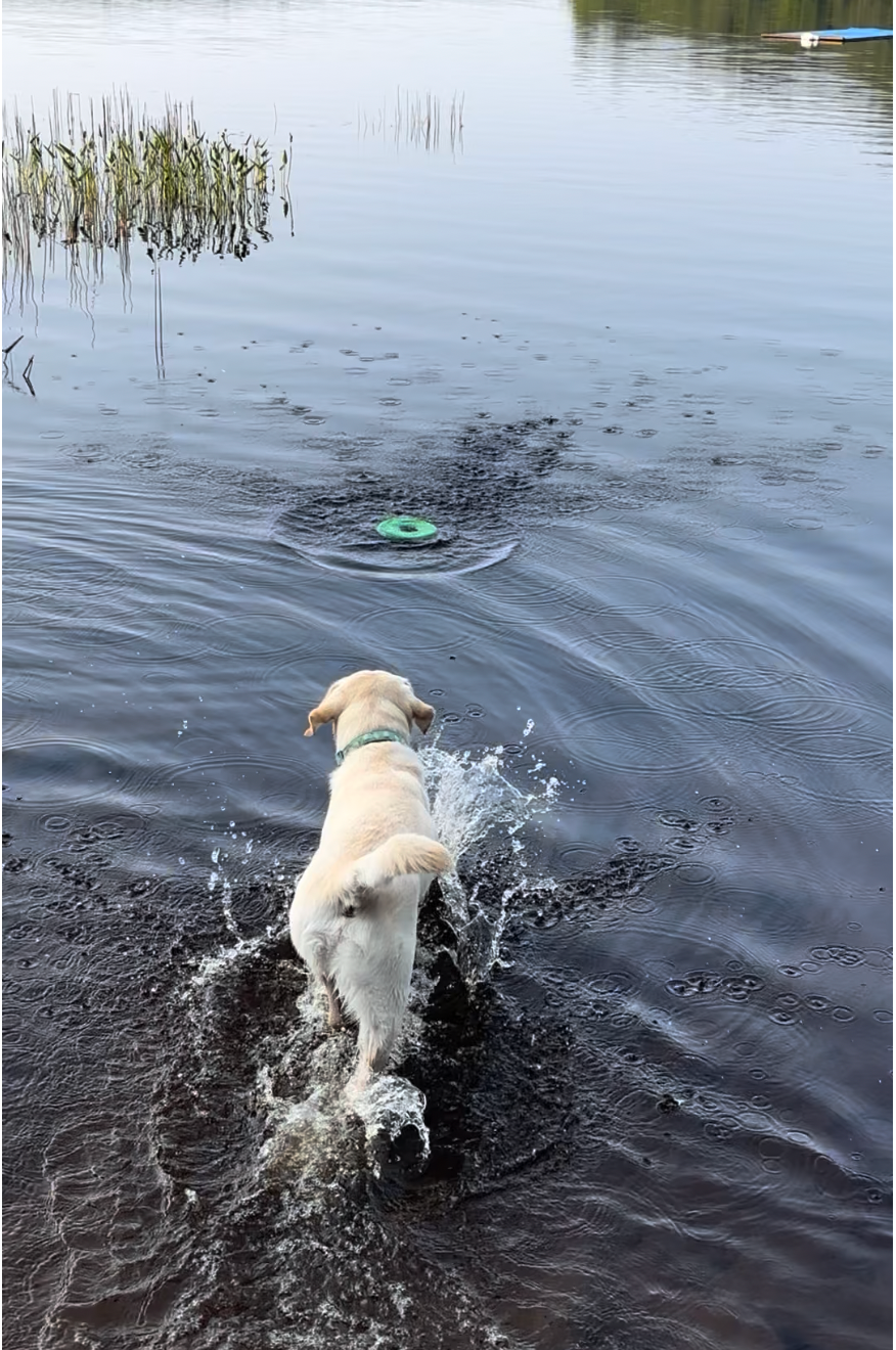
When we talk about vaccines of dogs*, we tend to split them into “core” and “non-core” vaccines.

(*The same applies to cats. I use dogs by default for posts like this, which sometimes gets me an earful, but I’m not actually ignoring cats.)

Core vaccines are those that every animal should get (e.g. rabies vaccine in areas where rabies exists, canine parvovirus in areas where dogs exist). Non-core vaccines are those that aren’t required by every dog, or that are less convincingly needed in every case.

Non-core vaccines are also often referred to as “lifestyle vaccines,” because the nature of the dog’s (or cat’s) lifestyle can put the animal at more or less risk of exposure to a disease, which affects the relative need for vaccination. Respiratory diseases are a great example. All dogs are at some degree of risk, but the risk is much higher in dogs whose lifestyles create more dog-dog contact (e.g. going to daycare, boarding, off-leash dog parks). That’s a good way to think about how to prioritize vaccination for an individual dog, but it misses a big part of the disease prevention equation.

When I’m assessing the need for vaccination in a pet, I think about two main things:

  1. Risk of exposure. The lifestyle aspect covers this.
  2. Risk of serious disease. This often gets ignored.

Some dogs are at higher risk of severe disease or death from respiratory infections. I’d put senior dogs, brachycephalics (i.e. flat-faced breeds), pregnant dogs, dogs with pre-existing heart or lung disease and dogs with compromised immune systems on that list. I’m more motivated to protect them because the implications of infection are higher, even if their risk of exposure may be fairly low.

Take my two dogs as an example (again):

Ozzie is 1.5 years old and healthy. If he gets a respiratory infection, most likely he’ll have transient disease and, while it will be annoying (for him and us) and I’d like to prevent it, odds are quite low he’ll suffer any serious consequences.

In contrast, Merlin is an 11 year old dog with chronic lymphoid leukemia who’s been getting chemotherapy for about 2 years. He’s doing really well, but he has a significant chronic disease and he’s old. If he gets a respiratory infection he’s at much greater risk of dying than Ozzie.

If we look at lifestyle of these two dogs, they’re similar, since they do everything together. The exception is in the summer when we go to a cottage for 2 weeks. Since 2 weeks with Ozzie at a cottage isn’t much of a vacation for us or Merlin, he went to a local day care for part of the time. (An exhausted Ozzie is a good Ozzie, and he often came home close to comatose, which was perfect.) So Ozzie has a major additional lifestyle risk factor, therefore he’ll get a respiratory vaccine again this summer (both because of the risk and because the day care requires it).

Merlin doesn’t have that same direct exposure risk, but he has some added risk through being exposed to Ozzie. Should he get a respiratory vaccine? If we just look at his lifestyle, we’d say no, he’s pretty low risk for exposure. However, his higher risk for severe disease increases my motivation to vaccinate him, and he’ll likely get a respiratory vaccine this summer at the same time Ozzie does.

Lifestyle is definitely important to consider, but we need to make sure we don’t just focus on the dog’s lifestyle and consider the dog (or cat) as a whole.

I debated writing about this now since it’s an ongoing situation with a very unclear outcome, but that’s medicine. We also don’t have a lot of camelid content on the blog, and there’s an infectious disease component to this case, so figured it was worthwhile.

Mickey (photo right) is an older male alpaca, part of my small group.

Why do I have alpacas? Here’s the short version:

  • I had a flock of rare breed sheep.
  • Coyotes starting picking off the sheep so I got a llama (Dolly) for predator control.
  • Dolly sucked at her job, so I was soon left with no sheep and a llama.
  • I then got some alpacas to keep the llama company.

This past Saturday, we found Mickey down on his side and couldn’t get up. I had thought that he looked a little weak in the hind legs earlier that week and this was presumably a progression of that problem. He was bright, alert and a pretty textbook spinal disease case. Specifically, his signs were consistent with a spinal lesion between the 3rd thoracic and 3rd lumbar vertebrae (T3-L3), given the severity of his hind limb abnormalities but pretty normal front limbs.

The two leading causes for this condition in an alpaca are:

  1. Parasite migration through the spinal cord due to infection with the deer meningeal worm Parelaphostrongylus tenuis (P. tenuis).
  2. Spinal trauma. Mickey and the boys chase each other around at times and it’s been icy. I didn’t appreciate anything in his back that would suggest a spinal fracture or other traumatic event, but sometimes it can be hard to tell.

While I couldn’t rule out trauma, parasitic migration made more sense, and P. tenuis is a major issue for camelids (like alpacas, llamas) and moose in some regions. Like many parasites, it has an unusual life cycle (see illustration below):

  • P. tenuis normally lives in white tailed deer, which are abundant around here. The adult parasites live in the subdural space and associated tissues in the central nervous system. Females lay eggs in blood vessels, and the eggs travel through the blood to the lungs where they develop into larvae (L1 stage). The larvae penetrate the small air sacs (alveoli) in the lungs and are coughed up and swallowed. They’re then passed in the feces, and subsequently infect slugs and snails, developing to their L3 larval stage.
  • If an animal ingests infected slugs or snails, the larvae move from the intestinal tract and somehow find their way to the central nervous system. In white tailed deer (the definitive host), they develop in gray matter of the spinal cord and then migrate to the subdural space, mature to adults and the life cycle starts again, usually without causing any problems for the deer. However, in other species like alpacas, the larvae don’t mature, and instead stay in the gray matter, causing inflammation and significant damage. Usually this damage predominantly affects control of the hind limbs, but depending on the location(s) to which the parasites migrate, damage to other parts of the spinal cord or brain can occur, so it’s on my list for any neurological disease in a camelid.

Back to Mickey. Given the odds of parasitic disease, the lack of any additional treatment I could possibly providefor trauma and the limitations of field imaging (e.g. x-rays), I’ve been treating him as a presumed P. tenuis larva migrans case with antiparasitics, anti-inflammatories and nursing care. I could have done a spinal tap to try to confirm my suspicion, but if it was unremarkable I still wouldn’t rule out P. tenuis, so I’d still treat him. Given the hassles of performing a spinal tap and the limited likelihood that I’d find a different cause, I didn’t bother. (If he was in hospital where it was easier and more convenient, I’d likely have done it, as it would be interesting to know the results.)

Drugs are part of the treatment for this disease, but nursing care is a huge component. Any down large animal is always a concern, as they can damage their muscles and nerves lying down for too long simply because of their weight. Fortunately, alpacas are much lower risk for complications from short term recumbency given their smaller size. However, I still need to keep him in a well bedded area and move him around so that he’s changing positions and not laying in wet (urine soaked) bedding. That also helps reduce the risk of pressure sores, another major concern in recumbent animals.

Over the past few days, he’s been up and down (figuratively and literally). Most days, I can get him up with some assistance and he’ll take a few steps with me supporting and steering him by holding his tail. Some days have been better than others.

With any neurological disease, the first thing I want to see is that the animal stops deteriorating. If the animal just keeps getting worse, the prognosis is really bad. Mickey plateaued pretty early, which was a good start.

Yesterday, I tried him in an ad hoc sling I made from a repurposed hammock. It wasn’t much of a success since he didn’t try to stand. A sling can be good to get an animal off their feet and let them use their limbs, with support. But Mickey didn’t use the sling like that, he just hung there, so there was a risk the sling would just cause more damage. It was a nice try, but I abandoned it, at least for now.

This morning, Mickey got up with assistance and took the most steps he’s made so far, walking from the barn to a run-in shed/barn. Movement is good, but walking on ice is bad. However, there was a pretty good path for him and the shed is well bedded, so I let him lead the way and that was his spot for a while.

The video below is from this afternoon – his strongest effort yet, but still with some pretty obvious major abnormalities.

What’s the prognosis for Mickey?

I keep waffling on that. Milestone #1 was when he stopped deteriorating. Now we need to see how he improves. Neurological damage is slow to improve, and the degree of improvement is hard to predict. As long as he keeps improving, there’s hope. Once that initial improvement plateaus, I don’t expect major improvement after that. Animals with neurological damage can still improve a bit at that stage, probably in part by learning how to compensate for their deficits, but we don’t expect to see a dramatic improvement later on.

I’d like to think he’s still in the improvement phase. It’s hit and miss; he’ll look good one time and then bad a little later… that’s life managing neurological disease. But he’s showing enough improvement and is otherwise stable enough that he’s worth treating. The video from today is by far the best he’s been so that’s encouraging.

He didn’t like me much before and he’s definitely not a fan now given all the poking, prodding and medicating, but that’s life with livestock. More updates to come (good or bad).

Life cycle figure from:

As awareness of canine infectious respiratory disease complex (CIRDC, formerly known as “kennel cough”) has spiked recently, there are more discussions happening about respiratory vaccines in dogs. A large number of different bacteria and viruses play a role in CIRDC. We can vaccinate against a few of them including parainfluenza virus (the most commonly diagnosed contributor to CIRDC), the bacterium Bordetella bronchiseptica (typically number 2 or 3 on the list of diagnosed contributors), canine adenovirus (pretty uncommon) and canine influenza (very sporadic).

We also have different ways to vaccinate dogs, specifically use of injectable versus mucosal (oral or intranasal) vaccines.

  • Injectable vaccines tend to induce a better systemic antibody responses. Mucosal vaccines provide a better local immune response at the mucosal surface. For respiratory infections, the local immune response is probably the most effective. There’s reasonable evidence that mucosal vaccines are superior to injectable vaccines for Bordetella. We don’t have good data for parainfluenza, but I’d assume the same applies. (We only have injectable influenza vaccines for dogs.)
  • Mucosal vaccines are modified live organisms – versions of Bordetella and parainfluenza that are still alive (i.e. functional) but have been attenuated so that while they can elicit an immune response, there is negligible risk of causing disease in the animal. We never say a modified live vaccine (MLV) is 100% guaranteed not to cause disease, but the risk is really low, and the protection is really good, so overall they’re beneficial for vaccination in “normal” animals. However, we tend to avoid MLVs in immunocompromised animals because low virulence organisms might be more likely to cause disease in an individual with a compromised immune system.

That’s the dog side. But, we have to remember that each dog is attached to one or more people too. When we vaccinate a dog with a mucosal vaccine, it sheds the modified bacterium/virus for a while, and might have a large load of the vaccine strain in their nose or mouth right after the initial administration.

That means people can be exposed to the vaccine strains as well. Generally, that’s not a big deal, and it’s really only a potential issue for Bordetella (because canine parainfluenza and canine adenovirus of any form don’t infect people). I get asked about this a lot, by both veterinarians and pet owners, and I write a similar post to this one every few years, but each time we have a bit more data.

Why is there concern about human exposure to Bordetella in canine vaccines?

  • Bordetella bronchiseptica can cause infections in people. They are rare, but they occur. So, if the “normal” Bordetella bronchiseptica can cause disease in people, we have to think about whether the vaccine strains can cause disease too.
  • The answer is “yes,” with a big “but” (actually, a series of “buts”).

Yes, there have been a couple of reports of human infections with canine vaccine-strain Bordetella, some of which are more convincing that others.

A recent report (Kraai et al. 2023) described vaccine-strain Bordetella bronchiseptica infection in a 43-year-old woman who was taking immunosuppressive medication.

  • She developed bronchitis with malaise and a mild fever two weeks after her dog had received an intranasal vaccine.
  • Bordetella bronchiseptica was isolated from her sputum. When it’s gene sequence was assessed, it was consistent with the vaccine strain.
  • She had mild disease and responded to antimicrobial treatment.

Clearly there is some risk with human exposure, that’s certain. Some groups have said to avoid MLVs in animals living with immunocompromised people. But let’s thing about that critically for a moment. All vaccination decisions require consideration of the costs (risks) versus benefits:

  • The risk to humans from canine vaccines is really low. Millions of doses of mucosal vaccines are given to dogs every year, yet human infections are still extremely rare.
  • Disease that has been reported in people who do get sick is mild.
  • Mucosal vaccination is superior to parenteral vaccination, and prevention of disease in dogs can also reduce the risk of exposure to the “wild type” (non-attenuated) strains of Bordetella in humans.

Broad “don’t use modified live vaccines in animals owned by high risk people” statements overlook a few big-picture issues:

  • The big one is the vaccine strain is much less likely to cause disease than the circulating (non-attenuated, disease causing) strains. A person is much more likely to be infected with the Bordetella from a naturally infected dog than from a vaccinated dog, so I’d rather prevent the dog from getting infected by vaccinating with the most effective method available.
  • Natural Bordetella infection (unlike vaccination) also tends to make the dog cough, which increases human exposure to any number of bugs in the dog’s respiratory tract.
  • If that dog needs treatment with antimicrobials, we run the risk of the person being exposed to antimicrobial resistant bacteria, some of which can pose additional risks to people.
  • Antimicrobials also increase the risk of the dog developing diarrhea, which can greatly increase human exposure to disease-causing bacteria in feces (especially if the dog poops on the floor).

Some more food for thought:

If I have a dog that was recently vaccinated with a mucosal vaccine, and I was asked to rank the top 5 zoonotic pathogens that are in the dog, vaccine-strain Bordetella wouldn’t even crack that list. There’s a mix of potentially disease-causing bacteria in every dog, all the time. Getting tunnel vision about one in particular, especially one that’s really quite low risk, is not helpful.

What about killed, injectable Bordetella vaccines?

Injectable killed Bordetella vaccines (which contain no live organisms, as the name suggested) do work, they just don’t work as well. If there’s significant concern from the owner, or some other unusual circumstance that makes use of a mucosal vaccine undesirable, then by all means, use an injectable vaccine. I’d consider that to be a rare situation.

Also bear in mind that killed “kennel cough” vaccines are just for Bordetella. They don’t include anything for parainfluenza virus, the most common cause of CIRDC. Parainfluenza is part of common combination “core” vaccines (e.g. DA2PP), but those vaccines don’t do a great job of protecting against paraflu. So, while an injectable Bordetella vaccine removes the risk of exposure to vaccine-strain Bordetella, it offers less protection against Bordetella and none against paraflu, so we have greater risk of disease in the dog overall, and the implications described above that come with it.

Let’s be clear: There’s never a zero risk situation when it comes to exposure to infectious bugs (from vaccination or pet ownership in general). We have to consider the risks and benefits in every situation.

But, almost always, for high risk households, I support vaccination whenever the dog’s lifestyle and risk factors indicate that Bordetella vaccination is warranted. I’d stick with mucosal vaccines for respiratory diseases whenever possible, since they provide much better protection and we can easily mitigate the very low risk from the vaccine. Those mitigation measures include:

  • Keeping the owner outside of the exam room when the dog is vaccinated.
  • Wiping the dog’s nose/mouth after vaccination to remove any major external contamination.
  • Recommending that the owner avoid direct contact with the dog’s oral and nasal secretions. That’s particularly important for the first 24 hours after vaccination, but it’s something I’d recommend for a high risk owner to always avoid.
  • Being diligent about routine hygiene practices (e.g. handwashing), especially after contact with the dog’s face (again, something that’s actually always important for a high risk owner).

Photo credit: Dr. Kate Armstrong (from Weese & Evason, Infectious Diseases of the Dog and Cat, A Color Handbook)

In the first two parts of this series, I explained a lot of the changes that have been made to the CLSI veterinary antimicrobial susceptibility testing guidelines, specifically those related to staphylococci and Enterobacterales (which includes E. coli and friends).  There’s less to say about Pseudomonas, but these changes will impact our use of the already limited range of available antimicrobials for this vexing bacterial genus.

New antimicrobial susceptibility breakpoints for Pseudomonas

Most veterinarians don’t realize that we don’t have established, species-specific breakpoints for many bug/drug combinations. The breakpoints veterinary labs use to call a bug susceptible or resistant to an antimicrobial are often extrapolated from other species and/or drugs. That probably works reasonably well most of the time, but not all the time. Species-specific breakpoints that are based on an understanding of the drug pharmacokinetics in that species are needed to have more confidence in the antimicrobial susceptibility testing results.

Breakpoints for enrofloxacin and marbofloxacin are now available for Pseudomonas isolates specifically from dogs. Previously, labs presumably chose a breakpoint for these isolates based on human fluoroquinolone breakpoints, the canine levofloxacin breakpoints, or the feline enrofloxacin breakpoints.  All of those are higher than the new canine breakpoints for these drugs. As a result, some bacterial isolates that would have previously been reported as susceptible will now be reported (more accurately) as resistant. This will seemingly reduce treatment options in some cases, but it’s actually a good thing, because we can have more confidence using these drugs for infections that are reported as susceptible, and should have fewer treatment failures.

New susceptible, dose dependent (SDD) breakpoints for Pseudomonas in dogs

This new breakpoint classification approach has been implemented for Pseudomonas and enrofloxacin and marbofloxacin. It’s based on recognition that we can often safely use higher doses of these drugs (at least in dogs), which we can use to overcome some degree of resistance:

Canine breakpoints for enrofloxacin and Pseudomonas

CategorySusceptible (5 mg/kg)SDD: 10 mg/kgSDD: 20 mg/kgResistant
MIC<0.06 ug/ml0.120.25> 0.5
  • If the MIC is >0.5 ug/mL, the bug is resistant. Don’t use this drug.
  • If the MIC is 0.25 ug/mL, the bug should be susceptible if we use a dose of 20 mg/kg
  • If the MIC is 0.12 ug/mL, the bug should be susceptible if we use a dose of >10 mg/kg
  • If the MIC is <0.06 ug/mL, the bug is susceptible at a dose of >5 mg/kg (although in dogs, I’d rather not go below 10 mg/kg, and in cats, I basically never use enrofloxacin because of safety issues)

If the MIC is <0.5 ug/mL, I’d only treat at 20 mg/kg. The bug might be susceptible to a lower dose, but we don’t know.

If the MIC is reported as <4, <2, <1 or <0.5 ug/mL, we’re screwed. We need to know that the bug has an MIC or no greater than 0.25 ug/mL in order for treatment with enrofloxacin to be effective, and none of those MICs tell us that clearly – the bacterium could still be susceptible or resistant. I would not use enrofloxacin in these cases.

Canine breakpoints for marbofloxacin and Pseudomonas (pretty similar)

CategorySusceptible (2.75 mg/kg)SDD: 5.5 mg/kgResistant
Minimum inhibitory concentration (MIC)<0.120.25> 0.5
  • If the MIC is >0.5 ug/mL, the bug is resistant. Don’t use this drug.
  • If the MIC is 0.25 ug/mL, the bug should be susceptible if we use a dose of 5.5 mg/kg
  • If the MIC is <0.125 ug/mL, the bug should be susceptible at a dose of >2.75 mg/kg

If the MIC is <0.5 ug/mL, only treat using a dose of 5.5 mg/kg. The bug might be susceptible to 2.75 mg/kg, but we don’t know.

If the MIC is reported as <4, <2, or <1 ug/mL, we don’t know if the bug is actually susceptible or resistant, so I would not use marbofloxacin.

There are also no disk diffusion breakpoints for these drugs and Pseudomonas, so we need the MIC data (which is based on broth microdilution) to determine susceptibility.

These changes will be disruptive but are important, because under the old guidelines labs are reporting a lot of bugs as susceptible when they really aren’t, or when higher drug doses are needed to treat effectively. It will take time for labs to implement the changes to their testing and reporting. In the interim, we need to look at the actual MIC, not just whether the lab classified the bug as susceptible or resistant under the old guidelines. Since labs may not test a wide range of drug concentrations, we’re going to have situations where we can’t properly interpret the results, at least until the labs make the necessary changes.

The Clinical and Laboratory Standards Institute (CLSI) has updated their main veterinary testing standards document: VET01SEd7E Performance Standards for Antimicrobial Disk and Dilution Susceptibility Tests for Bacteria Isolated From Animals, 7th Edition. Check out earlier posts for an overview of the relevant changes, and more specifics about the standards for staphylococci and chloramphenicol.

Today’s topic is about fluoroquinolones, a drug class with which I have a love-hate relationship. They’re great drugs, I just wish we’d massively decrease how much we use them, so they can keep being great drugs for animals and people for a long time. If we’re going to use a drug class like fluoroquinolones, we need to use it right. The news CLSI guidance will help with that, once we figure out how to get it actually rolled out.

There are three major changes in the latest update regarding fluoroquinolones:

  • changes in breakpoints for susceptible, intermediate and resistant bugs
  • introduction of dose-dependent breakpoints
  • removal of disk diffusion breakpoints

Changes in breakpoints

The mean inhibitory concentration (MIC) breakpoints for fluoroquinolones have been lowered, meaning some bacteria that would have been reported as susceptible using the older breakpoints will now be reported as resistant. Fluoroquinolones will therefore be an option in fewer cases, but when they are an option (i.e. when a bug is reported as susceptible), we should be more confident that they will be effective.

Susceptible, dose dependent (SDD) breakpoints

A major change in this update of the lab standards is the introduction of susceptibility reporting that is based on dose. Since we can safely increase the dose of fluoroquinolones quite a bit in most situations (enrofloxacin in cats being the main exception, and probably also any fluoroquinolone in very young animals), it makes sense to consider the dose when determining susceptibility. If we can get 2X or 4X the amount of drug at the site of infection, we can sometimes treat bacteria that would not have been inhibited with normal dosing.

A quick rundown is below.

Here’s a quick comparison of the changes regarding enrofloxacin for staphylococci and Enterobacterales in dogs:

GuidelineSusceptibleIntermediateSDD 10 mg/kgSDD 20 mg/kgResistant
6th edition< 0.5 ug/ml1-2  > 4
7th edition<0.06 ug/ml 0.120.25> 0.5

As with chloramphenicol, we have to figure out how to deal with this now, as labs make the necessary changes to their testing and reporting protocols.

  • A big barrier to this is the limited range of drug concentrations that most labs test. If the main commercial plates used by the labs don’t test low enough concentrations of the antimicrobials, we can’t interpret the results easily using the new guidelines.

In the interim, we need to look at the MIC values on the lab reports, not just where it says “S,” “I” or “R” (since S, I and R may be based on the old guidelines).

For enrofloxacin:

  • If the MIC is >0.5 ug/mL, the bug is resistant. Don’t use this drug.
  • If the MIC is 0.25 ug/mL, the bug should be susceptible if we use a dose of 20 mg/kg.
  • If the MIC is 0.12 ug/mL, the bug should be susceptible if we use a dose of >10 mg/kg.
  • If the MIC is <0.06 ug/mL, it’s susceptible at >5 mg/kg (although in dogs, I’d rather not go below 10 mg/kg and in cats, I basically never use enrofloxacin because of safety issues).

However, if the lab reports MICs of <4, <2, <1 or <0.5 ug/ml, you can see we’re kind of screwed. We need to know that the bug has an MIC of no greater than 0.25 ug/mL, and none of those higher MIC breakpoints tell us that clearly. For any of those, the bacterium could be susceptible, susceptible but only at a higher dose, or resistant. I would not use enrofloxacin in those cases if I can’t tell what, if any dose, will be effective.

Marbofloxacin is pretty similar. The new breakpoints are lower, and they’ve incorporated one susceptible, dose-dependent (SDD) breakpoint. They also removed the recommendations for disk diffusion testing (one of the other methods for antimicrobial susceptibility testing, vs broth microdilution).

GuidelineSusceptibleIntermediateSDD 5.5 mg/kgResistant
6th edition< 12 > 4
7th edition<0.125 0.25> 0.5
  • If the MIC is >0.5 ug/mL, the bugs is resistant. Don’t use this drug.
  • If the MIC is 0.25 ug/mL, the bug should be susceptible if we use a dose of 5.5 mg/kg.
  • If the MIC is <0.125 ug/mL, the bug should be susceptible at a dose of >2.75 mg/kg.

If the lab reports MICs of <4, <2, <1 or <0.5 ug/mL, we don’t know if if the bug is actually susceptible or resistant (so I’d stay away from this drug to be safe).

What about cats?

For cats, the breakpoints have changed as for dogs, but there are no susceptible dose dependent (SDD) breakpoints. Presumably that’s because we don’t have enough data to determine SDD breakpoints for cats. For enrofloxacin, it’s a non-issue for me, since I’d never use 10 or 20 mg/kg of this drug in a cat. For marbofloxacin, it’s a reasonable consideration since we can safely use higher doses. I wonder whether we’ll see labs report things for cats as they do for dogs. It’s not unreasonable, it just has less supporting data.

Yesterday, I wrote a post about a new version of CLSI’s Performance Standards for Antimicrobial Disk and Dilution Susceptibility Tests for Bacteria Isolated From Animals. There are some major changes in this update, and I sympathize with the diagnostic labs that now have to update their testing and reporting. It won’t happen overnight, because some may need to develop different testing panels to account for some of the different breakpoints, and they’ll need to change their reporting systems.

While we want to push labs to update their methods and reporting ASAP, we still have to manage patients in the interim. My next few posts will explain how I’ll be looking at interpretation of antimicrobial susceptibility testing results in the meantime.

I’ll start with chloramphenicol and staphylococci in dogs. Our typical methicillin-resistant Staphylococcus pseudintermedius (MRSP) tends to be resistant to multiple antimicrobial classes beyond beta lactams, and therefor has limited treatment options, so chloramphenicol susceptibility often gets included in reports for this bug.

The old breakpoints for chloramphenicol and staphylococci in dogs were:

  • Susceptible < 8 ug/mL
  • Intermediate 16 ug/mL
  • Resistant > 32 ug/mL

The *new* breakpoints for chloramphenicol and staphylococci in dogs are:

  • Susceptible < 2 ug/mL
  • Intermediate 4 ug/mL
  • Resistant > 8 ug/mL

I don’t think the intermediate category means much here, since chloramphenicol isn’t a drug for which we can safely increase the dose. So, practically speaking, the bacterium must have an MIC < 2 ug/mL for chloramphenicol to have a chance at being effective.

One big problem the labs will have is that at least one of the plates commonly used for susceptible testing of bacterial isolates from dogs won’t be able to differentiate susceptible from resistant to chloramphenicol based on these new guidelines, because the lowest concentration it contains is 8 ug/mL of chloramphenicol.

  • If there’s no growth in the 8 ug/mL well, then the bug was inhibited at that concentration and we know the MIC is 8 ug/mL or lower; but we won’t know if the true MIC is 8 ug/mL (resistant), 4 ug/mL (intermediate) or < 2 ug/mL (susceptible).
  • If the bacterium grows in the 8 ug/mL well, then the MIC is clearly greater than 8 ug/mL, meaning the bug will be clinically resistant to chloramphenicol.

Labs will need new plates with (at a minimum) 1, 2, 4 and 8 ug/mL chloramphenicol wells.

So, what do we do in the interim? We have to look at lab reports to see if an MIC is reported.

If your lab reports MICs:

  • Look at the MIC, not just S/I/R category.
  • If the MIC is 4-8 ug/mL, the bug is resistant, even if the report still lists it as “susceptible.”
  • If the MIC is “<8 ug/mL” or “<4 ug/mL,” we have no way to tell if the bug is resistant or susceptible. Since a large percentage of S. pseudintermedius from dogs will have MICs that fall in the 4-8 ug/mL range, odds are pretty good that treatment will fail if all we know if that the MIC is <8 ug/mL, so I’d consider it resistant to be on the safe side (see examples below).

If your lab does not report MICs:

  • I’d consider the bug resistant to chloramphenicol, since most staphylococci are resistant to this drug.
  • Ask your lab to start reporting MICs. As we learn more and get better guidelines, MICs are becoming more useful, and therefore important to include on antimicrobial susceptibility reports.

Diagnostic testing is a cornerstone of veterinary medicine that helps us optimize patient care, but there’s a lot of science behind it that people often forget. We collect a sample, send it off for testing and magically get the results, often without putting a lot of thought into what happens at the lab. Labs (should) follow standard guidelines for performing tests of all kinds that are developed to help ensure they provide relevant and accurate results. As with most things (medicine or otherwise), guidance changes over time as we learn more and as new issues are identified. Keeping up with those changes is critical.

The Clinical and Laboratory Standards Institute (CLSI) is an organization that sets standards for microbiology testing in humans and animals. The standards are updated regularly, with some updates being more impactful than others. Hot off the presses (just today!) is the latest update to the main veterinary testing standards document: VET01SEd7E Performance Standards for Antimicrobial Disk and Dilution Susceptibility Tests for Bacteria Isolated From Animals, 7th Edition. This is a major update with some changes that will have significant impacts on how lab results are reported and interpreted, including changes to some of the interpretive breakpoints for susceptibility of certain bacteria to specific antimicrobials.

A core part of antimicrobial susceptibility testing is determining the minimum inhibitory concentration (MIC) of an antimicrobial for a particular bacterial isolate. The MIC always needs to be interpreted in the context of each patient in the context of how much of the antimicrobial we can expect to get at the site of infection when the animal is treated. A bug could be susceptible to low concentrations of a drug (low MIC), but if drug levels we get in the infected tissue are even lower, it might not be effective. Conversely, a bug might has a fairly high MIC, but if the drug reaches the site of infection at even higher levels, it might still work.

Breakpoints consider how much drug is required to inhibit the bacterium and how high the concentration of drug is expected to reach in the body. The breakpoint is the MIC where things tip between susceptible and resistant (or susceptible, intermediate and resistant).

New “susceptible, dose dependent” (SDD) category

One major change in the updated lab standards is that they now provide guidance for a susceptible, dose dependent (SDD) category for certain bug/drug combinations. This goes beyond the familiar susceptible, intermediate or resistant classification. The SDD category considers the potential to use higher drug doses and how that impacts the bacterium’s susceptibility. That’s relevant for some drugs where we can use higher (but still reasonable and safe) doses.  

Take for example the guidance regarding enrofloxacin and both staphylococci and Enterobacterales (i.e. E. coli and relatives) in dogs.

The previous version of the lab standards said:

  • MIC <0.5 ug/mL: susceptible
  • MIC 1-2 ug/mL: intermediate
  • MIC >/=4 ug/mL: resistant

Two things have now changed. One is that the breakpoints have been lowered a bit. The other is that they’ve come up with dose-dependent interpretive breakpoints:

  • MIC <0.06 ug/ml: susceptible at a dose of 5 mg/kg once a day (FYI I pretty much never use a dose that low for enrofloxacin because of concerns about efficacy and development of resistance)
  • MIC 0.12 ug/ml: susceptible at a dose of 10 mg/kg once a day
  • MIC 0.25 ug/ml: susceptible at a dose of 20 mg/kg/day
  • MIC >/=0.5 ug/ml: resistant

The lower breakpoints mean we’ll see a lot more of these bugs reported as resistant to enrofloxacin. The dose-specific guidance will help figure out when we can still use enrofloxacin to treat some of these middle-ground MIC bugs, and hopefully reduce ineffective use of this important antimicrobial.

Communicating this to veterinarians will be a challenge, since it’s a completely new concept for most.

Other breakpoint changes

Some other breakpoints have changed in addition to the reduction of the enrofloxacin breakpoints mentioned above. One is the reduction of the susceptible breakpoint for chloramphenicol and staphylococci in dogs from <8 ug/ml to <2 ug/ml. Bacteria with MICs of 4-8 ug/ml previously would have been called susceptible to chloramphenicol, but now will be reported as resistant. That’s relevant because there are a lot of staphylococci with MICs in that range, and we now consider them non-susceptible to this drug. So, we’ll see a lot more reported chloramphenicol resistance.

More species-specific breakpoints

The updated standards have also added numerous species-specific breakpoints. Unfortunately we don’t have breakpoints for all bug/drug combinations in every species. In these cases, we either have no guidance or we extrapolate from humans or other animals (better than nothing, but not ideal since there can sometimes be big differences between species). Over time, as more data become available, we eventually get to the point where we can make more specific recommendations.

Urinary breakpoints

Another subtle but important change is modification of how site-specific breakpoints are reported. Most of these are based on serum drug levels, since that’s what should be present throughout most of the body, and we tend to have the most data about serum levels. However, we know that some drugs are excreted at high levels in urine, and those high levels can make bugs that are resistant to a drug in serum susceptible to it in urine.

For that reason, there are separate serum and urine breakpoints for some drug/bug combinations (e.g. E coli and amoxicillin). Urine breakpoints are higher because more drug ends up in the urine and will be able to inhibit some bacteria that would tolerate the lower concentrations of drug in other parts of the body. Previously, this was called a “UTI” (urinary tract infection) breakpoint. The problem was urine levels are only relevant for lower urinary tract infections (cystitis), not upper urinary tract infections (e.g. pyelonephritis). For an infection in the kidney, we need to rely on the drug that’s in serum, not in urine, so the UTI terminology has lead to confusion and misreporting. Now it’s clearer as the the updated standards call those breakpoints “(lower) urinary tract (ur).“ Hopefully that will result in fewer pyelonephritis samples being interpreted with urine breakpoints and leading to suboptimal antimicrobial treatment decisions.

Labs vary in how quickly they make changes when these standards are updated. Some are on the ball right away. Some are really slow. Some never adopt all the guidelines. (I saw a lab report yesterday from the US using an old guidance that was changed 10 years ago, leading to a bad treatment choice.) Veterinarians asking labs if/when they’re going to adopt the changes may help spur them on. It’s a hassle for labs to make the changes and communicate why things are now different in the lab reports, but it’s important to get this done for obvious reasons.

I spend a lot of time talking about antimicrobial misconceptions and dogmas. They are a big issue, because they often lead to unnecessary or excessive duration of antimicrobial use, use of more invasive routes of administration (e.g. intravenous over oral), or use of higher-tier antimicrobials than necessary.

I’ll just address one of these misconceptions today: Doxycycline should be avoided in young growing animals because of teeth staining and bone development.

Nope.

The concern about using doxycycline in young animals relates to calcium binding. Tetracycline and some of the older drugs in this class have substantial calcium binding and use of some tetracyclines has been shown to impact the development of teeth in various species, including dogs. It’s even sometimes used in wildlife rabies vaccine baits as a way to help determine if an animal ingested a bait when it was younger, because the tetracycline will cause detectable staining of the teeth.

Doxycycline is different. It doesn’t bind calcium to the same degree as other drugs in this class, so we can’t assume that what happens with other tetracyclines also applies to doxycycline.

Staining of teeth is a major concern in humans, so we can look there for some guidance. They’ve had the same dogma (i.e. avoid doxycycline in kids) for decades, but it’s been challenged and efforts are underway to refute it.

  • A review and commentary about kids and malaria stated, “It is time to rehabilitate doxycycline and to recommend it for malaria treatment in children under 8 years of age” because it is “a cheap drug with a broad therapeutic spectrum and very little evidence of serious adverse events.”
  • The American Academy of Pediatrics states “Although data are limited, evidence does not support dental staining as a consequence of doxycycline use in young children.” They recommend doxycycline for a variety of conditions in young kids, particularly tickborne diseases.

So, while we’re lacking data for dogs, information from other species shows that we shouldn’t be concerned about staining teeth or related effects in young animals, and we should therefore use doxycycline when it’s indicated even in young patients, as it is a lower-tier, safe and effective antimicrobial.

Unsubstantiated (or clearly disproven) dogmas persist in medicine (human and veterinary) and they’re really hard to address. However, we have to try to address them because they can cause harm.

This may be my last update on this topic in the short term (unless things change, of course).

The good news:

The bad news:

  • Well, it’s not really bad news, but we still don’t know what actually happened. That’s far from surprising, because with waxing and waning endemic disease conditions like CIRDC, we rarely have a clear picture of what happened and why.
  • Everything for me continues to point to a gradual increase in the rate of infectious respiratory disease in dogs over the past couple of years, with the usual intermittent local and regional peaks and valleys. This year’s issues were probably somewhat higher peaks overlaid on a higher baseline, making the issue more obvious and drawing more attention.

What was the cause of the increased cases of CIRDC?

I’m sticking to “the usual suspects, doing their usual thing, just at higher rates.” There’s been a lot of investigation looking for new pathogens and, as far as I know, nothing convincing has come to light. Given the number and quality of the research groups that have been looking, it’s pretty convincing that we don’t have a specific new pathogen that’s caused an outbreak of disease in dogs across North America.

What do we do now?

As with any outbreak, we try to learn some things from the experience:

1. Surveillance

This situation was a reminder that we don’t have a good surveillance system in place for CIRDC (or most other companion animal diseases). There’s no easy fix for that, especially with no money, so we need to continue to try to leverage the information that is available to better understand disease patterns. We need to do that on an ongoing basis, not just when there’s concern about increased cases, because when we’re concerned about an outbreak, we need to know the normal rates of disease to put things into context.

2. Vaccination

While we only have vaccines for a few of the important causes of CIRDC (Bordatella, parainfluenza, influenza), they are good vaccines, and we need to optimize their use in dogs that have a reasonable risk of exposure and/or a higher risk for severe disease.

3. Thinking about severe disease

The risk of severe disease in some dogs during outbreaks doesn’t get as much attention as it should; hopefully we’re changing that. In most dogs, CIRDC is a short term, self-limiting problem that’s fairly mild, just like upper respiratory infections in people. However, some dogs get really sick, and some even die. We can’t predict every dog that will have a severe outcome, but we know that there are groups that are at higher risk, particularly older dogs, dogs with pre-existing heart or lung disease, and brachycephalic dogs (i.e. flat-faced breeds). We need to think about minimizing exposure and maximizing vaccine coverage in these groups.

We also need to get people thinking about function over appearance in dogs they are breeding and buying. There is currently a disturbingly high number of anatomically disastrous dogs out there, because people have bred reasonably-functional brachycephalic breeds into extremely flat-faced dysfunctional dogs that have myriad respiratory issues, even without infectious diseases to complicate the situation (see the pictures below). As the French bulldog has shot to the top of the list of the most common breeds in the US, we’re going to see more dogs die from respiratory disease. Not all Frenchies are a mess, but there are enough of them that we see infectious and non-infectious complications in them all the time.

The recent situation with CIRDC also might get people thinking more about their dogs’ social networks and risks, and how to minimize those while having limited impacts on important or enjoyable aspects of dog ownership. We’ll be doing some work on dog social networks later this year, so stay tuned.

Image from: https://pedigreedogsexposed.blogspot.com/2017/08/the-frenchies-that-win-by-nose.html

test

I’ll take a break from writing about widespread canine infectious respiratory disease complex (CIRDC) in North America to talk about a single case of a rare disease in a dog. Wageningen Veterinary Research has reported a case of Bluetongue infection in a dog in the Netherlands, a disease of significant consequence to livestock that’s recently been found again in the country.

Bluetongue is a viral disease caused by the unoriginally named bluetongue virus (BTV), which is endemic in many parts of the world, especially tropical and subtropical countries. However, a lot of countries put in significant effort to control this virus and maintain disease-free status, because it has major impacts on food animals. The infection often kills sheep, and while it doesn’t usually kill cattle, infection can cause a major drop in milk production. A single case of bluetongue in a previously disease-free country can result major livestock export restrictions. The Netherlands managed to control bluetongue since their last outbreak in 2007, but it was found again in the country earlier this year; based on the strain, it’s suspected the virus may have been imported from Italy.  

The bluetongue-affected dog in this report was a 3.5-year-old pregnant dog from a Dutch dairy farm. The dog had severe signs of illness, including shortness of breath, pulmonary edema, severe emaciation and lethargy. It’s interesting that someone considered bluetongue and tested the dog, especially since the disease wasn’t known to be present in cattle on the farm, though it was subsequently identified in two cattle after the dog’s diagnosis. Thanks to an astute vet and access to testing, a diagnosis of bluetongue was made through detection of BTV by PCR. Not surprisingly, it was the same strain of BTV that’s been circulating in sheep and cattle in the Netherlands.

There’s no mention of whether the dog survived and/or if it was treated. For something like this, we’d be focusing on supportive care and treating the consequences of the infection, while the dog hopefully fought off the virus itself, since we don’t have a specific antiviral treatment for BTV (or at least not one that we know is safe and effective in a dog).

This isn’t the first case of bluetongue in a dog, but it’s a very rare diagnosis. Interestingly, most of the reported canine cases have been in pregnant dogs. We don’t know why that is, but it could be for a number of potential reasons, or a combination thereof, including:

  • Just a coincidence
  • Greater likelihood of testing a sick pregnant dog
  • Increased susceptibility of dogs to infection during pregnancy

How was the dog infected with BTV?

Livestock-to-livestock transmission of BTV is mainly by biting midges (little insects), so the virus can move with infected sheep and cattle, infected midges (blown off course by storms or hitching a ride in a vehicle), or contaminated raw food items that somehow end up being fed to livestock (e.g. food scraps).

How this dog was infected isn’t clear. Contact with or ingestion of colostrum, fetal fluids or fetal membranes, raw meat or blood from an infected ruminant are considered the most likely routes of transmission. However, spread by infected midges can’t be ruled out. This dog lived on a dairy farm, had direct contact with cattle and their environment, and may have had access to high-risk tissues like placentas, so there were a variety of potential sources of exposure.

Does/did this dog pose a risk to other animals?

Presumably not. It’s assumed that dogs are “dead end hosts” for BTV. Dead end hosts can have serious disease but usually don’t produce or excrete enough virus to pass infection on to others. But that is an assumption here, and especially with a rare infection like BTV in a dog, we can’t have complete confidence in assumptions.

Does this case change anything in the big picture?

Probably not much. It’s a reminder of the unpredictability of infectious diseases, the need to consider the whole human/animal/environment ecosystem (versus having tunnel vision about certain species), and that spillover of infections from the main host to others probably occurs a lot more than we recognize for a wide range of infectious diseases.